AI to Assist in Classifying Lung Cancer and Diseases (including COVID-19)
Tomasz Bednarz on March 22, 2020
The evolution of GPGPU (General Purpose Graphics Processing Units) as compute accelerators opened up new possibilities in High-Performance Computing space (Hagoort, 2019). Modern platforms enable to apply super heavy workloads for not only accelerating and scaling up Machine Learning, Deep Learning and Artificial Intelligence algorithms, but also to solve problems such as weather simulations, fluid flow simulations, cyclones predictions, computer vision problems for autonomous vehicles support, image processing and analysis. GPUs (Graphics Processing Units) are designed with focus on the arithmetic throughputs especially with thousands of threads running at the same time, make them so very powerful as computational tools (Bednarz, 2010).
In Medicine, and especially looking at the classification of lung disease, decision on metrics and level of progression depends on medical practitioner and their years of experience. AI is mature enough today, to assist in making quicker and more accurate recommendations, that often can save lives.
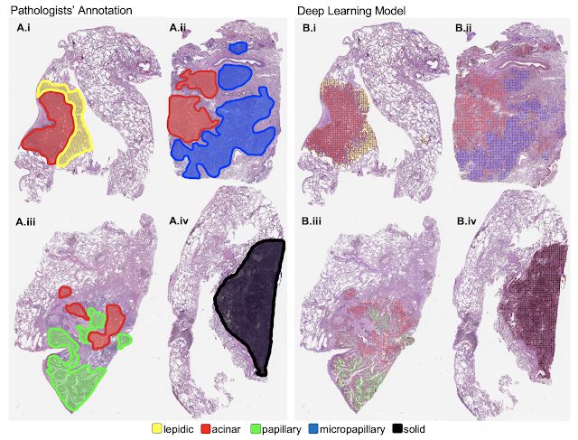
AI can help in classifying lung cancer at the pathologist level (Wei, et. al 2019; NVIDIA,2019). For instance having 143 images annotated by the pathologist, they could use them to train neural networks and achieve evaluation metrics for the model within 95% confidence intervals of agreement with the pathologist’s assessment, which is great.

Imaging, AI and Radiomics can also help in understanding and fight against COVID-19 (Quibim, 2020). As per today, we know there is no effective cure for the virus yet, but looking at CORVID-19 virus and progressions of its influence on lungs can help to understand the impact better, and hopefully be used as one of the input for testing various drugs.
The recent project proposal my team and collaborators at UNSW are developing is around using High Resolution Computed Tomography for Early Detection of Lung Disease. High-resolution computed tomography (HRCT) is likely to supersede chest x-rays as the primary tool for the diagnosis of lung diseases and has great potential to be used in widespread (particularly industry specific) health surveillance programs. In order to be meaningfully and reliably used, there is large scope for these computationally intensive scans to be analyzed remotely using computational platforms accessible through virtual laboratories (Bednarz, 2014) which can feature various artificial intelligence (AI), image analysis, image processing and visual analytics tools that can be further automated and scaled up through workflow management frameworks. Connecting high-performance computational frameworks with massive imaging data collected allow us to develop deep-learning-based training algorithms that could be applied to automatically in-paint and detect abnormal changes in the lungs. Use of intuitive visualization-based reporting system has the potential to provide detailed innovative and rapid reporting capability for medical practitioners.
There are lots of business opportunities in that space, use AI for Good!
PS. For everyone who is interested in AI and its applications to various fields, I strongly recommend you to have a look at NVIDIA GTC conference: https://www.nvidia.com/gtc/.
References
Bednarz, T. (2010). Introduction to OpenCL. OpenCL Workshop Brisbane. Available at https://www.slideshare.net/TomaszBednarz1/introduction-to-opencl-2010.
Bednarz, T. (2014). Image Analysis and Processing for everyone. Available at http://cloudimaging.net.au/.
Bednarz, T., Wang, D., et al. (2014). Cloud Based Toolbox for Image Analysis, Processing and Reconstruction Tasks. Signal and Image Analysis for Biomedical and Life Sciences. Available at https://link.springer.com/chapter/10.1007/978-3-319-10984-8_11.
Hagoort, N. (2019). Exploring the GPU Architecture. NIELS HAGOORT. Available at: https://nielshagoort.com/2019/03/12/exploring-the-gpu-architecture/.
NVIDIA (2019). AI Helps Classify Lung Cancer at the Pathologist Level. NVIDIA News Center. Available at https://news.developer.nvidia.com/ai-helps-classify-lung-cancer-at-the-pathologist-level/.
Quibim (2020). Imaging, AI and Radiomics to Understand and Fight Coronavirus CORVID-19. Available at https://quibim.com/2020/02/14/imaging-ai-and-radiomics-to-understand-and-fight-coronavirus-covid-19/.
Wei, J.W., Tafe, L.J., Linnik, Y.A., Vaickus, L.J., Tomita, N. and Hassanpour, S. (2019). Pathologist-level classification of histologic patterns on resected lung adenocarcinoma slides with deep neural networks. Nature Scientific Reports, 9, Article number: 3358. Available at https://www.nature.com/articles/s41598-019-40041-7.